Перейти к содержимому


Фото

Shatter Cones Of The Ladoga Impact Volcanic Structure. Macro And Micro Scales.


  • Чтобы отвечать, сперва войдите на форум
4 ответов в теме

#1 В.Юрковец

В.Юрковец

    Advanced Member

  • Administrators
  • 1 417 сообщений

Опубликовано 15 Май 2015 - 20:50

Only two of distinct shatter cone been found on Ladoga yet. They are took from the seabed in the coastal area of Cape Vladimirovskiy. This is the west coast of Lake Ladoga. Here, uplifted rim comes to the surface (see. Geological map of Ladoga IVS - sampling point "a"). Shatter cones were formed in schistose rock - quartz-sericite slate, which affected the structure of both the macroscopic and microscopic scales. Below are photos of the shatter cones and areas of thin concussion, which is clearly visible fracture and specific shock metamorphism of shatter cones.


ShM_2.JPG.jpg


A third small shatter cone was found to the south of the Ladoga impact crater. Its origin is probably associated with falling of the Ladoga's satellite cosmic body.


#2 В.Юрковец

В.Юрковец

    Advanced Member

  • Administrators
  • 1 417 сообщений

Опубликовано 15 Май 2015 - 21:40

SHATTER CONES IN A ROCKS


Im_Shutter_cone_4.JPG.jpg

Shatter cone in a slate with a cut of which a thin section across of the shock fracture was made.


Im_Shutter_cone_5.JPG.jpg

Shatter cone (view from the apex) in a slate with a cut of which a thin section along of the shock fracture was made.


Im_shatter_cone_6.JPG.jpg

Shatter cone in a slate with a perfectly circular apex (diameter approximately 50 mm).


Im_shatter_cone_7.JPG.jpg

View "from below".


Im_Shatter_cone_8.jpg

Im_Shatter_cone_9.jpg

Shatter cone in "eucrite".


#3 В.Юрковец

В.Юрковец

    Advanced Member

  • Administrators
  • 1 417 сообщений

Опубликовано 15 Май 2015 - 22:00

SHATTER FRACTURES IN THIN SECTIONS


Im_Shutter_cone_10.jpg

Im_Shutter_cone_11.jpgT

Thin section 4i (transverse). The shatter fractures. Microdisplacements expressed in the peculiar character of extinction. Field width 0.28 mm. Plane polarized light (top) and cross nicols (bottom).


Im_Shutter_cone_12.jpg

Im_Shutter_cone_13.jpg

The same is.


Im_Shutter_cone_14.jpg

Im_Shutter_cone_15.jpg

The same is. At the top right kink bands in biotite, indicating an impact, are noticeable.


#4 В.Юрковец

В.Юрковец

    Advanced Member

  • Administrators
  • 1 417 сообщений

Опубликовано 21 Май 2015 - 20:35

There's also a sample of allogeneic breccia (Vendian fragment of quartz sandstone) was sampled. In his grains structure of planar fracture were detected. They exactly reproduce the morphology of shatter cones of macroscopic level.


SHATTER CONES IN A QUARTZ GRAINS

( Russian - http://dna-genealogy...-макроуровень/)


Im._Shutter_cone_1.jpg

Thin section 7. Plane polarized light. Field width 0,28 mm. Quartz grain.


Im._Shutter_cone_2.jpg

Thin section 7. Plane polarized light. Field width 0,28 mm. Other quartz grain.


Im._Shutter_cone_2_a.JPG.jpg

Thin section 7. Plane polarized light. Field width 0,28 mm. Drawing.


Im_Shatter_cone_upright_arrow_1.JPG.jpg

Thin section 7. Plane polarized light. Field width 0,28 mm. Hiatuses showing the formation of the tips.


#5 В.Юрковец

В.Юрковец

    Advanced Member

  • Administrators
  • 1 417 сообщений

Опубликовано 22 Май 2015 - 13:43

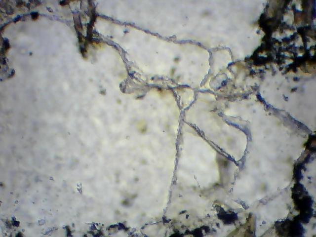
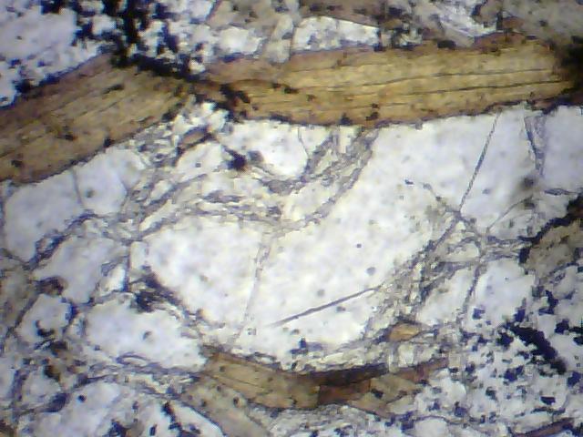
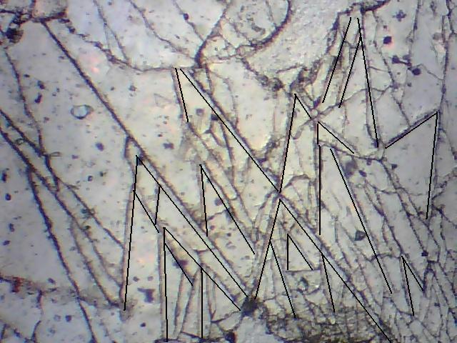
For example. In the bottom is represented shatter cone (branch) of micron scale


Shatter_cones_micron_scale.JPG.jpg


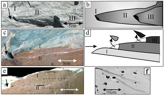
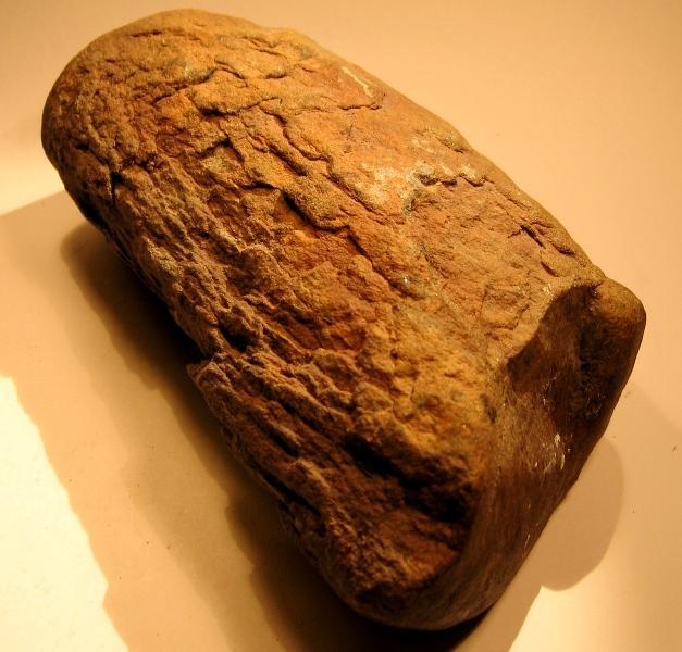
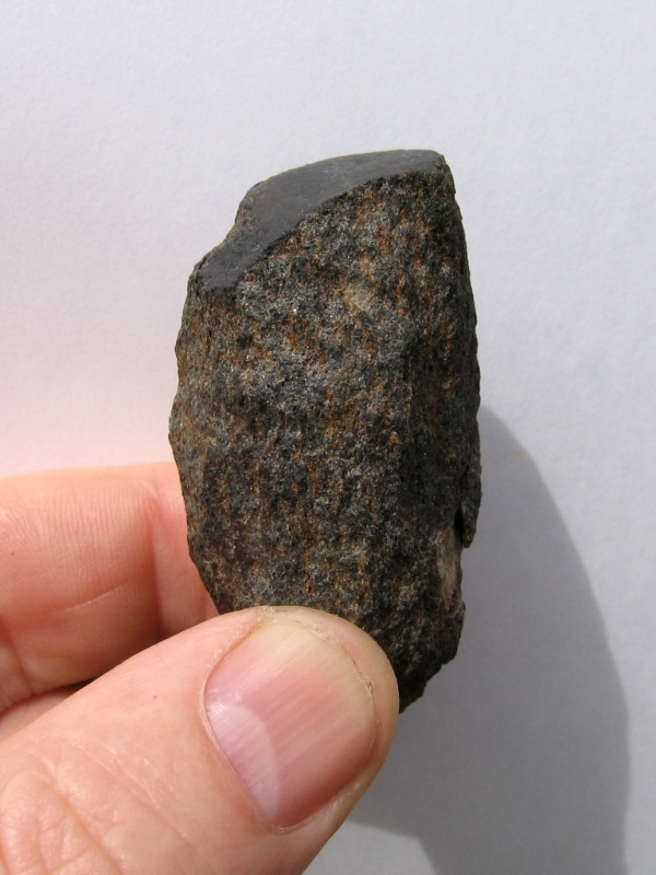
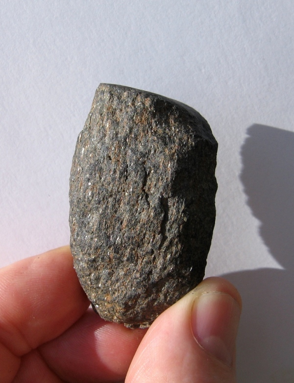
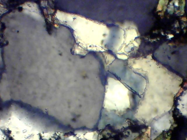
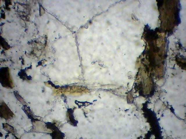
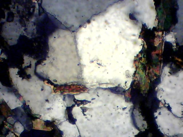
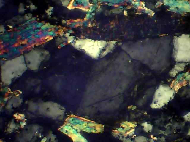
Fig. 4. The external and internal structure of shatter cones in dolomite samples from Kentland quarry. Photograph (a) and schematic map (b ) of a surface with three aligned shatter cones (marked I, II, and III) in hierarchic, cone-on-cone order. © Three-dimensional view of a shatter cone. A front sawcut reveals an internal surface, marked II, that branched away from the upper, external surface, marked I. The branching point is marked by an arrow and the branched surface has a characteristic spoon-like shape. The internal surface II is striated in the same direction as I (framed), and the two surfaces become nearly parallel a few centimeters from the branching point. The dotted lines delineate the cone tip, which commonly breaks off exposed samples. The overall structure explains 3D horse-tail structures. (d) A schematic cross section describing the internal structure of (a), (b ) and ©. (e) A shatter cone cross-section displaying a spoon-like branch which bifurcated from a point (arrow) on its parent surface. Further branching (framed) occurs from II to generate an additional spoon-like branch with a similar shape.Note the inverse curvature direction of branched surfaces; I is convex downward and II is concave upward (see text). (f) SEM image displaying multiple branching at micron scales. Note that cone tips are preserved in the internal branches presented in both (e) and (f).


Reference: Amir Sagy, Jay Fineberg, Ze’ev Reches. SHATTER CONES: BRANCHED, RAPID FRACTURES FORMED BY SHOCK IMPACT. Journal of Geophysical Research 109: B10209. doi:10.1029/2004JB003016.


0 пользователей читают эту тему

0 пользователей, 0 гостей, 0 скрытых

Copyright © 2024 Академия ДНК-генеалогии. Климатический филиал